Showing posts with label parkinsons. Show all posts
Showing posts with label parkinsons. Show all posts
Wednesday, February 26, 2014

depression and PD
Although depression is the most common psychiatric comorbidity in PD
[43], it is often underdiagnosed or undertreated [44]
43 Lemke MR, Fuchs G, Gemende I, et al. Depression
and Parkinson's disease. J Neurol. 2004;251 Suppl
6:VI/24-7.
44 Jasinska-Myga B, Putzke JD, Wider C, et al. Depres-
sion in Parkinson's disease. Can J Neurol Sci.
2010;37:61–6.
The prevalence of depression in PD varies between 20 % and 40 % of PD
patients [49, 50]
Richard IH, McDermott MP, Kurlan R, et al. A ran-
domized, double-blind, placebo-controlled trial of
antidepressants in Parkinson disease. Neurology.
2012;78:1229–36.
The largest randomized controlled study in PD assessed the
efficacy of paroxetine and venlafaxine in PD. Both drugs
were superior to placebo in treating depression, but failed to
alleviate symptoms of anxiety.
Accuracy of the Microsoft Kinect sensor for measuring movement in people with Parkinson's disease
reposted from
http://predictpd.blogspot.ca/2014/02/accuracy-of-microsoft-kinect-sensor-for.html
http://predictpd.blogspot.ca/2014/02/accuracy-of-microsoft-kinect-sensor-for.html
Accuracy of the Microsoft Kinect sensor for measuring movement in people with Parkinson's disease
Gait Posture. 2014 Jan 22. pii: S0966-6362(14)00024-1. doi: 10.1016/j.gaitpost.2014.01.008. [Epub ahead of print]
Galna B, Barry G, Jackson D, Mhiripiri D, Olivier P, Rochester L.
Author information
- Institute for Ageing and Health, Newcastle University, Newcastle upon Tyne, United Kingdom.
- Culture Lab, School of Computing Science, Newcastle University, Newcastle upon Tyne, United Kingdom.
- Institute for Ageing and Health, Newcastle University, Newcastle upon Tyne, United Kingdom.
Abstract
BACKGROUND:

The Microsoft Kinect sensor (Kinect) is potentially a low-cost solution for clinical and home-based assessment of movement symptoms in people with Parkinson's disease (PD). The purpose of this study was to establish the accuracy of the Kinect in measuring clinically relevant movements in people with PD.
METHODS:
Nine people with PD and 10 controls performed a series of movements which were measured concurrently with a Vicon three-dimensional motion analysis system (gold-standard) and the Kinect. The movements included quiet standing, multidirectional reaching and stepping and walking on the spot, and the following items from the Unified Parkinson's Disease Rating Scale: hand clasping, finger tapping, foot, leg agility, chair rising and hand pronation. Outcomes included mean timing and range of motion across movement repetitions.
RESULTS:
The Kinect measured timing of movement repetitions very accurately (low bias, 95% limits of agreement <10% of the group mean, ICCs >0.9 and Pearson's r>0.9). However, the Kinect had varied success measuring spatial characteristics, ranging from excellent for gross movements such as sit-to-stand (ICC=.989) to very poor for fine movement such as hand clasping (ICC=.012). Despite this, results from the Kinect related strongly to those obtained with the Vicon system (Pearson's r>0.8) for most movements.
CONCLUSIONS:
The Kinect can accurately measure timing and gross spatial characteristics of clinically relevant movements but not with the same spatial accuracy for smaller movements, such as hand clasping.
Wednesday, February 19, 2014
FEEL THE RHYTHM, FEEL THE RHYME: NEW INSIGHTS INTO MUSICAL BEAT PERCEPTION
reposted from

FEEL THE RHYTHM, FEEL THE RHYME: NEW INSIGHTS INTO MUSICAL BEAT PERCEPTION
Music and rhythm are fundamental and essential components of human civilization. From traditional Papa New Guinean log-drums (some of which live on Stanford’s campus), to Indian tabla, to modern computer-generated sound design: musical rhythm reverberates across an incredibly diverse range of cultures and times. Even non-musicians experience music in complex ways. For instance, when we dance we anticipate beat placement to coordinate our hands, feet, arms, and legs, while also twirling our dance partners in sync with the music (or so we hope).
Our need for music has also materialized in the clinical world. It has been shown that patients with Parkinson’s disease, who often exhibit difficulty initiating motor movements, can move more fluidly when listening to music. These patients who struggled simply to walk are able to move freely with remarkable rhythm when listening to their favorite songs. While anecdotal, these impressive results suggest the dire need to better understand the nature of music’s influence over the brain, and possibly tailor music as a novel neurological therapy (ref 1).
Although musical cognition may seem trivial, our brains must process an enormous amount of auditory and motor information to engage with music and internalize its meter. How is it that we are able to ‘count’ the meter of a song and prepare our movements accordingly? What is it about steady meter that facilitates movement in patients with motor disabilities? Unfortunately, despite our common interactions with music, extremely little is known regarding how our brains process it.
Brain waves: what are they, and what do they do?
One reason for our lack of knowledge of musical processing stems from the technological limitations of recording from human brains. Two non-invasive techniques that can measure the activity of the brain are theelectroencephalogram (EEG) and magnetoencephalogram (MEG), which record electrical and magnetic signatures of neural activity, respectively. These techniques are useful when using rapidly changing stimuli such as music because they can record instantaneous electromagnetic changes in the brain. On the other hand, they are limited in their spatial resolution. A helpful analogy is to imagine an EEG or MEG electrode as a microphone hanging over a large stadium – the microphone will pick up a large roar after a touchdown, but cannot discern individual conversations.
Despite their limitations, these techniques enable us to record oscillating “waves” of activity that occur normally in our brains and which may give us insights into the brain activity underlying musical processing. Essentially, these brain waves are electromagnetic patterns that arise from the activity of our neurons and the communication between different regions in our brains. Various waves have characteristic frequencies and are associated with different processing tasks. In fact, these different wave frequencies are so common that they have been given identifying names using the greek alphabet (gamma, beta, delta, etc).
Two particular types of brain waves – beta (β: 15-20 Hz) and gamma (γ: 30-100 Hz) – may have implications for musical cognition. Past studies have found β waves are generally associated with active planning, thinking, and more specifically, motor planning, movement, and control. γ waves, on the other hand, are associated with deep concentration and have been observed to fluctuate with external visual and auditory sensory input. Given that these waves are correlated with muscular coordination and sensory stimuli, respectively, new theories have proposed that they are also involved in music and beat processing (ref 2,ref 3).

Might Beta and Gamma waves facilitate steady musical beat perception?
Takako Fujioka, a new professor at Stanford University, and her colleagues have discovered that β and γ waves are indeed important for beat perception. The researchers had non-musicians passively listen to sequences of steady beats while they recorded from the subjects’ auditory cortices (the region of the brain responsible for sound perception) using MEG. They discovered that β and γ waves precisely followed the beats with periods of low and high amplitudes. When they looked more closely, the researchers found notable differences in how the two types of brainwaves followed the rhythm of the beats. When the researchers occasionally omitted a beat during the sequence, they discovered that β remained at an elevated level but γ continued on its normal path, as if the beat had actually been heard. In other words, although both wave trajectories followed the rhythm of the beats, only γ was able to maintain the rhythm when a beat was skipped (ref 4, see figure). This suggests that as we listen to music, specific brain waves are associated with different aspects of the music – part of our brain seems to be listening carefully to the actual beats we hear, while other aspects of brain activity appear to be predicting the beats we expect to hear.
Moving again to the beat
These new findings on brain waves may give some insight behind symptomatic motor stiffness observed in many patients with Parkinson’s disease, and the fact that music sometimes allows these patients to move normally. Parkinson’s patients often have highly elevated β waves, which are associated with resistance to movement initiation. As mentioned, Dr. Fujioka and her team discovered that β waves decrease in amplitude immediately following each beat in a sequence. Perhaps music facilitates movement in these patients by affecting the underlying neurological processes behind their β waves in ways that decrease β wave amplitude and help initiate movement. Although there is still much more to investigate on this topic, these new findings are promising and have opened a door for future research in this field.
REFERENCES:
1: de Dreu, M. J., van der Wilk, A. S. D., Poppe, E., Kwakkel, G., and van Wegen, E. E. H. (2012). Rehabilitation, exercise therapy and music in patients with Parkinson’s disease: a meta-analysis of the effects of music-based movement therapy on walking ability, balance and quality of life. Parkinsonism & Related Disorders, Volume 18 (Supplement 1): S114-S119. doi: 10.1016/S1353-8020(11)70036-0.
2: Joundi, R.A., Jenkinson, N., Brittain, J.-S., Aziz, T. Z. and Brown, P. (2012). Driving Oscillatory Activity in the Human Cortex Enhances Motor Performance. Current Biology, Volume 22 (Issue 5): 403-407. doi: 10.1016/j.cub.2012.01.024.
3: Noda, T., Kanzaki, R., and Takahashi, H. (2013). Amplitude and phase-locking adaptation of neural oscillation in the rat auditory cortex in response to tone sequence. Neuroscience Research, available online 15 November 2013. doi: 10.1016/j.neures.2013.11.002.
4: Fujioka, T., Trainor, L. J., Large, E. W. and Ross, B. (2009), Beta and Gamma Rhythms in Human Auditory Cortex during Musical Beat Processing. Annals of the New York Academy of Sciences, 1169: 89–92. doi: 10.1111/j.1749-6632.2009.04779.x
Tuesday, February 18, 2014
Thursday, January 30, 2014
Parkinson gene: Nerve growth factor halts mitochondrial degeneration
reposted from
http://www.mpg.de/7871678/parkinson-gdnf
http://www.mpg.de/7871678/parkinson-gdnf
Medicine . Neurosciences
Parkinson gene: Nerve growth factor halts mitochondrial degeneration
New link discovered between processes associated with a Parkinson’s-related gene defect
January 30, 2014
Neurodegenerative diseases like Parkinson’s disease involve the death of thousands of neurons in the brain. Nerve growth factors produced by the body, such as GDNF, promote the survival of the neurons; however, clinical tests with GDNF have not yielded in any clear improvements. Scientists from the Max Planck Institute of Neurobiology in Martinsried and their colleagues have now succeeded in demonstrating that GDNF and its receptor Ret also promote the survival of mitochondria, the power plants of the cell. By activating the Ret receptor, the scientists were able to prevent in flies and human cell cultures the degeneration of mitochondria, which is caused by a gene defect related to Parkinson’s disease. This important new link could lead to the development of more refined GDNF therapies in the future.
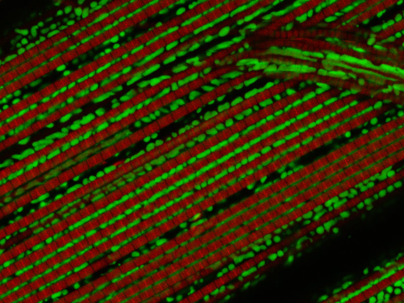
In his “Essay on the Shaking Palsy” of 1817, James Parkinson provided the first description of a disease that today affects almost 280,000 people in Germany. The most conspicuous symptom of Parkinson’s disease is a slow tremor, which is usually accompanied by an increasing lack of mobility and movement in the entire body. These symptoms are visible manifestations of a dramatic change that takes place in the brain: the death of large numbers of neurons in theSubstantia nigra of the midbrain.
Despite almost 200 years of research into Parkinson’s, its causes have not yet been fully explained. It appears to be certain that, in addition to environmental factors, genetic mutations also play a role in the emergence of the disease. A series of genes is now associated with Parkinson’s disease. One of these isPINK1, whose mutation causes mitochondrial dysfunction. Mitochondria are a cell’s power plants and without them, a cell cannot function properly or regenerate. Scientists from the Max Planck Institute of Neurobiology and their colleagues from Munich and Martinsried have now discovered a hitherto unknown link that counteracts mitochondrial dysfunction in the case of a PINK1 mutation.
The PINK1 gene emerged at a very early stage in evolutionary history and exists in a similar form for example in humans, mice and flies. In the fruit flyDrosophila, a mitochondrial defect triggered by a PINK1 mutation manifests in the fraying of the muscles. Less visible, the flies’ neurons also die. The scientists studied the molecular processes involved in these changes and discovered that the activation of the Ret receptor counteracts the muscle degeneration. “This is a really interesting finding which links the mitochondrial degeneration in Parkinson’s disease with nerve growth factors,” reports Rüdiger Klein, the head of the research study. Ret is not an unknown factor for the Martinsried-based neurobiologists: “We already succeeded in demonstrating a few years ago in mice that neurons without the Ret receptor die prematurely and in greater numbers with increasing age,” says Klein.
The Ret receptor is the cells’ docking site for the growth factor GDNF, which is produced by the body. Various studies carried out in previous years showed that the binding of GDNF to its Ret receptor can prevent the early death of neurons in the Substantia nigra. However, clinical studies on the influence of GDNF on the progression of Parkinson’s in patients did not lead to any clear improvement in their condition.
The new findings from basic research suggest that the mitochondrial metabolism is boosted or re-established through Ret/GNDF. “Based on this finding, existing therapies could be refined or tailored to specific patient groups,” hopes Pontus Klein, who conducted the study within the framework of his doctoral thesis. This hope does not appear to be completely unfounded: The scientists have already discovered a Ret/GDNF effect in human cells with aPINK1 defect similar to that observed in the fruit fly. It may therefore be possible to search for metabolic defects in the mitochondria of Parkinson’s patients in future. A specially tailored GDNF therapy could then provide a new therapeutic approach for patients who test positively.
SM/HR
Sunday, January 26, 2014
How does the brain create sequences? And how do separate small elements come together to become a unique and meaningful sequence?
reposted from
AAAS and EurekAlert! are not responsible for the accuracy of news releases posted to EurekAlert! by contributing institutions or for the use of any information through the EurekAlert! system.
PUBLIC RELEASE DATE:
26-Jan-2014
[ Close Window ]
Contact: Vitor Cunha
351-966-619-794
JLM&A, SA
How does the brain create sequences?
And how do separate small elements come together to become a unique and meaningful sequence?
When you learn how to play the piano, first you have to learn notes, scales and chords and only then will you be able to play a piece of music. The same principle applies to speech and to reading, where instead of scales you have to learn the alphabet and the rules of grammar.
But how do separate small elements come together to become a unique and meaningful sequence?
It has been shown that a specific area of the brain, the basal ganglia, is implicated in a mechanism called chunking, which allows the brain to efficiently organise memories and actions. Until now little was known about how this mechanism is implemented in the brain.
In an article published today (Jan 26th) in Nature Neuroscience, neuroscientist Rui Costa, and his postdoctoral fellow, Fatuel Tecuapetla, both working at the Champalimaud Neuroscience Programme (CNP) in Lisbon, Portugal, and Xin Jin, an investigator at the Salk Institute, in San Diego, USA, reveal that neurons in the basal ganglia can signal the concatenation of individual elements into a behavioural sequence.
"We trained mice to perform gradually faster sequences of lever presses, similar to a person who is learning to play a piano piece at an increasingly fast pace." explains Rui Costa. "By recording the neural activity in the basal ganglia during this task we found neurons that seem to treat a whole sequence of actions as a single behaviour."
The basal ganglia encompass two major pathways, the direct and the indirect pathways. The authors found that although activity in these pathways was similar during the initiation of movement, it was rather different during the execution of a behavioural sequence.
"The basal ganglia and these pathways are absolutely crucial for the execution of actions. These circuits are affected in neural disorders, such as Parkinson or Huntington's disease, in which learning of action sequences is impaired", adds Xin Jin.
The work published in this article "is just the beginning of the story", says Rui Costa. The Neurobiology of Action laboratory at the CNP, a group of around 20 researchers headed by Rui Costa, will continue to study the functional organisation of the basal ganglia during learning and execution of action sequences. Earlier this year, Rui Costa was awarded a 2 million euro Consolidation Grant by the European Research Council to study the mechanism of Chunking.
###
About Rui Costa
Dr. Costa received his D.V.M. from the Technical University of Lisbon in 1996. He entered the GABBA graduate program from University of Porto in 1997, and performed his Ph.D. studies with Dr. Alcino Silva at UCLA from 1998 to 2002 in the field of learning and memory, investigating the molecular and cellular mechanisms underlying the learning disabilities associated with NF1. For this work, Dr. Costa received the Young Investigator Award from the National Neurofibromatosis Foundation in 2001, and was a finalist of the Lindsley Prize from the Society for Neuroscience in 2003. He then joined Dr. Miguel Nicolelis at Duke University for his postdoctoral training, where he established multi-site neuronal recordings in behaving mice to investigate mechanisms of action generation and skill learning. Dr. Costa became a Section Chief at the National Institutes of Health in 2006. In 2009 he became an Investigator of the Champalimaud Neuroscience Programme, and received a Marie Curie International Reintegration Grant and a European Research Council Starting Grant. In 2010 he received the Seeds of Science Prize for Life Sciences, and in 2012 the Young Investigator Award from SFN.
About Xin Jin
Xin Jin got his BSc at China Agricultural University and his PhD from Shanghai Jiao Tong University, China. He then joined the lab headed by Rui Costa at the National Institutes of Health, where he studied the neural mechanisms underlying the parsing and concatenation of action sequences. He is currently an Assistant Professor at the Salk Institute in San Diego, California.
About Fatuel Tecuapetla
Fatuel Tecuapetla studied Biology in Puebla, Mexico and received his PhD from Universidade Nacional de Mexico. He came to CNP in 2009 and joined the Neurobiology of Action Lab, headed by Rui Costa where he worked on the role of basal ganglia on the initiation and performance of action sequences. He is currently starting his own group at the UNAM in Mexico City.
About the Champalimaud Neuroscience Programme (CNP)
The CNP is an international programme which strives to unravel the neural basis of behaviour. The concept of the programme takes into account the fact that basic neuroscience research can have a significant impact on the understanding of brain function, which in turn may contribute to the understanding and possible treatment of neurological and psychiatric illnesses.
Media
AAAS and EurekAlert! are not responsible for the accuracy of news releases posted to EurekAlert! by contributing institutions or for the use of any information through the EurekAlert! system.
HOME DISCLAIMER PRIVACY POLICY TERMS & CONDITIONS CONTACT US TOP
Copyright ©2014 by AAAS, the science society. |
Saturday, December 7, 2013
Parkinson's study at centre stage: NBS and Dancing with Parkinson's
reposted from http://www.rbnonline.ca/media/28264/


Parkinson's study at centre stage
Uploaded on 11/27/2013 4:00:21 PM
Production Credit(s)
Jessica Defreitas Reporter
Jacqueline Bradley Crew
Armen Bedakian Crew
Tags
No tag!
Embed Code
More From This Program


More From Special Feature


It’s a busy time of year for Canada’s National Ballet School. They aren’t only preparing for their annual Nutcracker performance. They have become the home of a new study for Parkinson’s.
Tuesday, November 19, 2013
Caffeine could help some Parkinson’s symptoms
reposted from here
Read more: http://www.ctvnews.ca/health/health-headlines/caffeine-could-help-some-parkinson-s-symptoms-1.900449#ixzz2l9wxOu7j
Read more: http://www.ctvnews.ca/health/health-headlines/caffeine-could-help-some-parkinson-s-symptoms-1.900449#ixzz2l9wnkhQ1
Caffeine could help some Parkinson’s symptoms

Dr. Ronald Postuma of McGill University Health Centre explains the double blind study suggests caffeine has an effect on motor skills.

Avis Favaro takes a look at a new study that finds caffeine to help people with Parkinson's move easier and more fluid.
Read more: http://www.ctvnews.ca/health/health-headlines/caffeine-could-help-some-parkinson-s-symptoms-1.900449#ixzz2l9wxOu7j
CTVNews.ca Staff
Published Wednesday, August 1, 2012 2:01PM EDT
Last Updated Wednesday, August 1, 2012 5:02PM EDT
Published Wednesday, August 1, 2012 2:01PM EDT
Last Updated Wednesday, August 1, 2012 5:02PM EDT
Drinking caffeine each day might help Parkinson’s patients move a little easier, making their walking more fluid and improving their quality of life, new Canadian research has found.
The researchers say the improvements are small, but their research suggests there’s something about caffeine that blocks malfunctioning brain signals in Parkinson’s patients.
The finding was a surprising one and came during a study to test caffeine as a treatment for daytime sleepiness among Parkinson’s patients. Though it didn’t do much for keeping fatigue at bay, it did seem to help with movement.
RELATED STORIES
PHOTOS
Bull Dog Coffee owner Stuart Ross displays a fancy cappuccino he created in Toronto on Tuesday, Sept. 23, 2008. (Nathan Denette / THE CANADIAN PRESS)
The study looked at 61 people with Parkinson’s disease with symptoms of daytime sleepiness as well as the tremors, rigidity and slow movement that marks the brain disease.
The volunteers were given either a placebo pill or a pill with 100 milligrams of caffeine, twice a day for three weeks. The dose was then bumped up to 200 milligrams twice a day for three weeks -- the equivalent of between two and four cups of coffee per day.
The researchers report in the journal Neurology that the caffeine didn’t really help improve sleepiness. But it did help with some Parkinson’s symptoms.
After the six weeks of study, the patients who had taken the caffeine saw an improvement of about five points in Parkinson’s severity ratings, compared to those who didn’t take the caffeine.
The caffeine group also averaged a three-point improvement in the speed of movement and amount of stiffness, compared to the placebo group.
Though the improvements were small, they were enough that patients noticed them, says study author Dr. Ronald Postuma, with the Research Institute of the McGill University Health Centre.
“It's a modest difference but it is real,” he told CTV News. “And it is there on objective measurements. Patients moved better while on caffeine.”
Parkinson’s patient Archie Christian says the disease can be like walking with heavy weights on your legs. But he says after taking caffeine pills as part in the study, he found he could walk faster and easier.
“In a few days, I noticed a big difference,” he says.
When he went off them, he became slower and stiffer.
Studies have already shown that people who get caffeine daily from coffee are less likely to develop Parkinson’s disease in the first place. This study suggests it may be that caffeine somehow blocks malfunctioning brain signals.
The study authors note that the number of patients they studied was small, and the length of their study was short. They say it’s also possible that the effects of caffeine may lessen over time.
Still, Dr. Edward Fon of the Parkinson Foundation of Canada says the study is intriguing.
“I think it warrants more investigation because this is relatively safe compared to other experimental mediations --safe and cheap compared to newer experimental medications,” Dr. Fon told CTV News.
Dr. Michael Schwarzschild, of Massachusetts General Hospital in Boston, who wrote an accompanying editorial, says he’s not ready to recommend caffeine for all Parkinson’s patients.
“Although the results do not suggest that caffeine should be used as a treatment in Parkinson’s disease, they can be taken into consideration when people with Parkinson’s are discussing their caffeine use with their neurologist,” he said in a statement.
The study was supported by the Canadian Institute of Health Research and the Webster Foundation.
With a report from CTV medical specialist Avis Favaro and producer Elizabeth St. Philip
Read more: http://www.ctvnews.ca/health/health-headlines/caffeine-could-help-some-parkinson-s-symptoms-1.900449#ixzz2l9wnkhQ1
Subscribe to:
Posts (Atom)