Brain’s immune cells linked to Alzheimer’s, Parkinson’s, schizophrenia
Salk and UC San Diego scientists conducted vast microglia survey, revealing links to neurodegenerative diseases and psychiatric illnesses
LA JOLLA—Scientists have, for the first time, characterized the molecular markers that make the brain’s front lines of immune defense—cells called microglia—unique. In the process, they discovered further evidence that microglia may play roles in a variety of neurodegenerative and psychiatric illnesses, including Alzheimer’s, Parkinson’s and Huntington’s diseases as well as schizophrenia, autism and depression.
“Microglia are the immune cells of the brain, but how they function in the human brain is not well understood,” says Rusty Gage, professor in Salk’s Laboratory of Genetics, the Vi and John Adler Chair for Research on Age-Related Neurodegenerative Disease, and a senior author of the new work. “Our work not only provides links to diseases but offers a jumping off point to better understand the basic biology of these cells.”
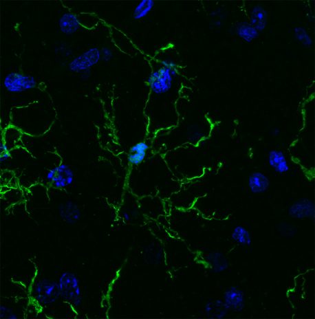
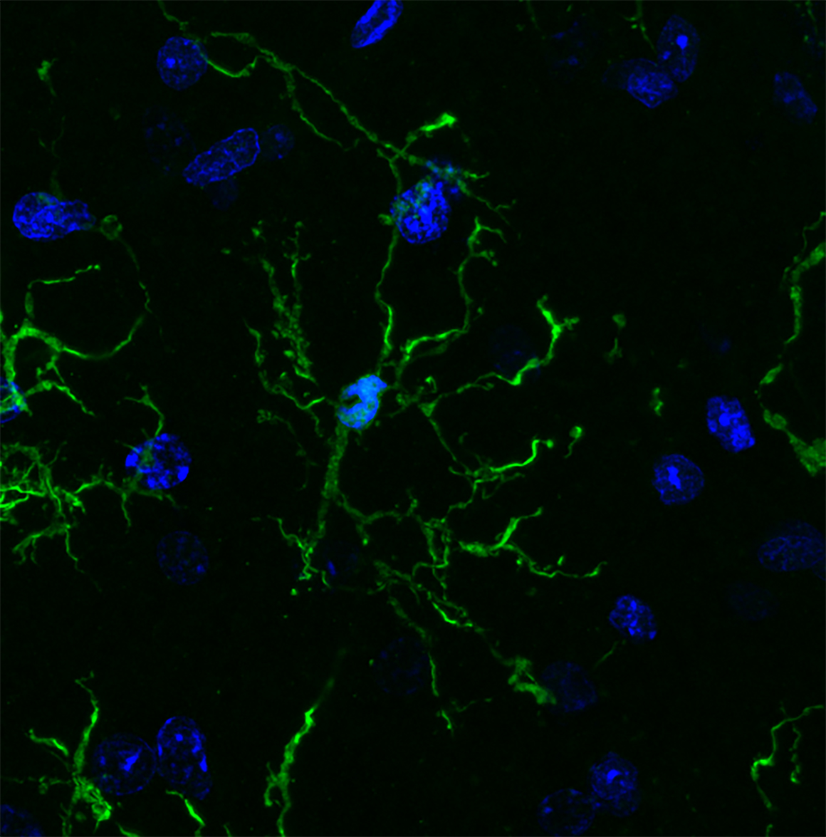
Salk and UC San Diego scientists conducted a vast survey of microglia (pictured here), revealing links to neurodegenerative diseases and psychiatric illnesses.
Genes that have previously been linked to neurological diseases are turned on at higher levels in microglia compared to other brain cells, the team reported in Science on May 25, 2017. While the link between microglia and a number of disorders has been explored in the past, the new study offers a molecular basis for this connection.
“These studies represent the first systematic effort to molecularly decode microglia,” says Christopher Glass, a Professor of Cellular and Molecular Medicine and Professor of Medicine at University of California San Diego, also senior author of the paper. “Our findings provide the foundations for understanding the underlying mechanisms that determine beneficial or pathological functions of these cells.”
Microglia are a type of macrophage, white blood cells found throughout the body that can destroy pathogens or other foreign materials. They’re known to be highly responsive to their surroundings and respond to changes in the brain by releasing pro-inflammatory or anti-inflammatory signals. They also prune back the connections between neurons when cells are damaged or diseased. But microglia are notoriously hard to study. They can’t be easily grown in a culture dish and quickly die outside of a living brain.
Nicole Coufal, a pediatric critical care doctor at UC San Diego, who also works in the Gage lab at Salk, wanted to make microglia from stem cells. But she realized there wasn’t any way to identify whether the resulting cells were truly microglia.
“There was not a unique marker that differentiated microglia from circulating macrophages in the rest of the body,” she says.
David Gosselin and Dylan Skola in the Glass lab, together with Coufal and their collaborators, set out to characterize the molecular characteristics of microglia. They worked with neurosurgeons at UC San Diego to collect brain tissue from 19 patients, all of who were having brain surgery for epilepsy, a brain tumor or a stroke. They isolated microglia from areas of tissue that were unaffected by disease, as well as from mouse brains, and then set out to study the cells. The work was made possible by a multidisciplinary collaboration between bench scientists, bioinformaticians and clinicians.
The team used a variety of molecular and biochemical tests—performed within hours of the cells being collected—to characterize which genes are turned on and off in microglia, how the DNA is marked up by regulatory molecules, and how these patterns change when the cells are cultured.
Microglia, they found, have hundreds of genes that are more highly expressed than other types of macrophages, as well as distinct patterns of gene expression compared to other types of brain cells. After the cells were cultured, however, the gene patterns of the microglia began to change. Within just six hours, more than 2,000 genes had their expression turned down by at least fourfold. The results underscore how dependent microglia are on their surroundings in the brain, and why researchers have struggled to culture them.
From left: Rusty Gage (Salk Institute) and Christopher Glass (UC San Diego).
Next, the researchers analyzed whether any of the genes that were upregulated in microglia compared to other cells had been previously implicated in disease. Genes linked to a variety of neurodegenerative and psychiatric diseases, they found, were highly expressed in microglia.
“A really high proportion of genes linked to multiple sclerosis, Parkinson’s and schizophrenia are much more highly expressed in microglia than the rest of the brain,” says Coufal. “That suggests there’s some kind of link between microglia and the diseases.”
For Alzheimer’s, more than half of the genes known to affect a person’s risk of developing the disease were expressed more highly in microglia than other brain cells.
In mice, however, many of the disease genes weren’t as highly expressed in microglia. “That tells us that maybe mice aren’t the best model organisms for some of these diseases,” Coufal says.
More work is needed to understand exactly how microglia may be altered in people with diseases, but the new molecular profile of microglia offers a way for researchers to begin trying to better culture the cells, or coax stem cells to develop into microglia for future studies.
Other researchers on the study were Baptiste Jaeger, Carolyn O’Connor, Conor Fitzpatrick, Monique Pena, and Amy Adair of the Salk Institute; Inge Holtman, Johannes Schlachetzki, Eniko Sajti, Martina Pasillas, David Gona, and Michael Levy of the University of California San Diego; and Richard Ransohoff of Biogen.
Altered circadian rhythm worsens Parkinson's disease, researchers show
Date:
April 5, 2016
Source:
Temple University Health System
Summary:
Chronic lack of sleep and irregular sleep-wake cycles may be risk factors of Parkinson's disease, new work suggests. In an animal model, the researchers show that disturbances in circadian rhythm that exist before Parkinson's onset dramatically worsen motor and learning deficits brought on by the disease.
Share:
FULL STORY
This is Domenico Praticò, MD, Professor in the Departments of Pharmacology and Microbiology and the Center for Translational Medicine at Lewis Katz School of Medicine at Temple University
Credit: Lewis Katz School of Medicine at Temple University
Chronic lack of sleep and irregular sleep-wake cycles may be risk factors of Parkinson's disease, new work by researchers at the Lewis Katz School of Medicine at Temple University (LKSOM) suggests. In an animal model, the researchers show that disturbances in circadian rhythm that exist before Parkinson's onset dramatically worsen motor and learning deficits brought on by the disease.
The new work, led by Domenico Praticò, MD, Professor in the Departments of Pharmacology and Microbiology and the Center for Translational Medicine at LKSOM, is the first to demonstrate that an environmental factor -- chronic daily exposure to long periods of light with brief exposure to dark, which alters circadian rhythm -- can exacerbate Parkinson's symptoms and pathology. The findings appear online April 5 in the journal Molecular Psychiatry.
Patients with Parkinson's disease often suffer from recurrent sleep disorders and disturbances in circadian rhythm, the roughly 24-hour biological cycle of humans. But whether those disturbances impact the development and progression of Parkinson's has been unclear. "Many think that sleep disturbances are secondary to Parkinson's disease," Dr. Praticò explained. "But circadian rhythm disturbances are increasingly reported before the onset of Parkinson's, suggesting that they could be risk factors."
After age 60, the majority of Parkinson's disease cases are idiopathic, their cause unknown. According to Dr. Praticò, it is probable that in those cases, the disease arises as a result of interactions between genes and environmental risk factors. The latter include chronic stress, sleep disorders, and circadian disturbances, all of which affect the function of the central nervous system, potentially contributing to the pathology that characterizes Parkinson's disease.
Dr. Praticò and colleagues investigated the role of altered circadian rhythm using a well-established mouse model of Parkinson's disease, in which treatment with MPTP, a neurotoxin, reproduces aspects of the disease in mice. The researchers divided animals into two groups.
The first, the control group, was maintained on a regular circadian schedule, being exposed to 12 hours of light followed by 12 hours of dark each day. In the second group, circadian rhythm was altered through daily exposure to 20 hours of light followed by just four hours of dark. After 60 days, some animals from each group were treated with MPTP.
Assessments of movement and behavior showed that all mice treated with MPTP developed Parkinson's disease, but animals with altered circadian rhythm experienced significant learning impairments. They also exhibited severe motor deficits, with drastic reductions in motor coordination and motor learning skills -- far worse than the deficits observed in MPTP-treated mice with normal circadian rhythm.
To understand why circadian rhythm disturbance worsens Parkinson's disease, Dr. Praticò and his team examined the brains of affected mice. In a region known as the substantia nigra, they observed significant reductions in neurons that produce dopamine, the loss of which is a major molecular feature of Parkinson's disease. "The substantia nigra is the epicenter of Parkinson's disease," Dr. Praticò said. "Cells normally die in that region of the brain, but our study shows that circadian rhythm disturbance accelerates cell death there."
In addition, cells known as microglia, which normally protect neurons, were superactive in circadian-disrupted MPTP-treated mice. The overactivation of microglia can actually worsen neuroinflammation and potentially speed the progression of Parkinson's disease.
The next challenge is to see if the findings can be replicated in other animal models. "If those studies are successful, we'll then try to reestablish normal circadian rhythm in circadian-disrupted animals to explore the possibility of reversing brain inflammation and cell death," Dr. Praticò said.
The outcomes of those studies could have important implications for the prevention and treatment of Parkinson's disease in persons with chronic sleep disorders.
E Lauretti, A Di Meco, S Merali, D Pratic�. Circadian rhythm dysfunction: a novel environmental risk factor for Parkinson’s disease. Molecular Psychiatry, 2016; DOI: 10.1038/MP.2016.47