Showing posts with label optogenetics. Show all posts

Friday, April 4, 2014

Light-activated neurons from stem cells restore function to paralysed muscles

resposted from


Light-activated neurons from stem cells restore function to paralysed muscles

4 April 2014
Diagram of optical muscle control system
A new way to artificially control muscles using light, with the potential to restore function to muscles paralysed by conditions such as motor neuron disease and spinal cord injury, has been developed by scientists at UCL and King’s College London.
The technique involves transplanting specially-designed motor neurons created from stem cells into injured nerve branches. These motor neurons are designed to react to pulses of blue light, allowing scientists to fine-tune muscle control by adjusting the intensity, duration and frequency of the light pulses.
In the study, published this week in Science, the team demonstrated the method in mice in which the nerves that supply muscles in the hind legs were injured. They showed that the transplanted stem cell-derived motor neurons grew along the injured nerves to connect successfully with the paralyzed muscles, which could then be controlled by pulses of blue light.
“Following the new procedure, we saw previously paralysed leg muscles start to function,” says Professor Linda Greensmith of the MRC Centre for Neuromuscular Diseases at UCL’s Institute of Neurology, who co-led the study. “This strategy has significant advantages over existing techniques that use electricity to stimulate nerves, which can be painful and often results in rapid muscle fatigue. Moreover, if the existing motor neurons are lost due to injury or disease, electrical stimulation of nerves is rendered useless as these too are lost.”
Muscles are normally controlled by motor neurons, specialized nerve cells within the brain and spinal cord. These neurons relay signals from the brain to muscles to bring about motor functions such as walking, standing and even breathing. However, motor neurons can become damaged in motor neuron disease or following spinal cord injuries, causing permanent loss of muscle function resulting in paralysis
“This new technique represents a means to restore the function of specific muscles following paralysing neurological injuries or disease,” explains Professor Greensmith. “Within the next five years or so, we hope to undertake the steps that are necessary to take this ground-breaking approach into human trials, potentially to develop treatments for patients with motor neuron disease, many of whom eventually lose the ability to breathe, as their diaphragm muscles gradually become paralysed. We eventually hope to use our method to create a sort of optical pacemaker for the diaphragm to keep these patients breathing.”
The light-responsive motor neurons that made the technique possible were created from stem cells by Dr Ivo Lieberam of the MRC Centre for Developmental Neurobiology, King’s College London.
“We custom-tailored embryonic stem cells so that motor neurons derived from them can function as part of the muscle pacemaker device.” says Dr Lieberam, who co-led the study. “First, we equipped the cells with a molecular light sensor. This enables us to control motor neurons with blue light flashes. We then built a survival gene into them, which helps the stem-cell motor neurons to stay alive when they are transplanted inside the injured nerve and allows them to grow to connect to muscle.”

Media contact: Harry Dayantis
Image: Diagram showing how the system works (credit: Barney Bryson)

Links:

Friday, November 8, 2013

HOW ALGAE COULD ONE DAY CURE PARKINSON’S DISEASE

reposted from:
http://www.fastcompany.com/3020560/reverse-engineered/how-algae-could-one-day-cure-parkinsons-disease


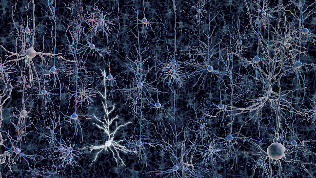
AN ACTIVATED NEURON IN A TANGLE OF NEURONS.


HOW ALGAE COULD ONE DAY CURE PARKINSON’S DISEASE

A LOOK AT OPTOGENETICS TECHNOLOGY'S JOURNEY FROM MURKY WATER TO RESEARCH LAB.
Ever see a green film form on a still pond? It happens because alga needs sunlight to complete the photosynthesis cycle, the process it uses to convert carbon dioxide and water into the sugar it "eats." It’s that drive to seek the light that makes Chlamydomonas reinhardtii, a single-celled alga, a brain research superstar and the core of optogenetics technology--which uses light-sensitive proteins to study how neurons employ electrochemical signals to produce memories, thoughts, and behaviors. It is helping scientists research brain diseases and disorders such as Parkinson's, schizophrenia, depression, stroke, PTSD, addiction, some forms of blindness, and more.
"The ideal way to study the property of different types of neurons is to control individual types of cells independently and see what happens when you alter one type of cell. Optogenetics helps to realize this goal," says Feng Zhang, core member at Broad Institute of Massachusetts Institute of Technology (MIT) and Harvard, who was part of the Stanford University team that created the technology. "Brain cells are normally not sensitive to light. So by introducing light-sensitive proteins into specific types of neurons, we can now selectively control that specific type of neuron by shining light in the brain."

THE DISCOVERIES

The Chlamydomonas proteins, called channelrhodopsins, were discovered on the alga's eyespot by a Texas Health Science Center research team in 2002. Led by John Spudich, director of the Center for Membrane Biology at the University of Texas-Houston Medical School, they found that the protein not only seeks light, but it also generates electric currents when exposed to it. Chlamydomonas then uses that energy to propel itself toward the light.
A subsequent 2003 paper from German researchers Georg Nagel, Ernst Bamberg, and Peter Hegemann showed that the channelrhodopsins weren’t just useful for photosynthesis, but they could also stimulate electrical currents in animal cells. And that was the big breakthrough Ed Boyden and his team were waiting for.

THE CONNECTION

Back in 2000, Boyden, who is currently an associate professor in the departments of biological engineering and brain and cognitive sciences at MIT, and his advisor at Stanford University Karl Deisseroth, had begun dreaming up ways to control and study neurons, the building blocks of the nervous system. Boyden had trained in electrical engineering and physics before pursing his PhD in neuroscience at Stanford, and he was fascinated with exploring the electrical activity of neurons. The pair's thoughts kept turning to using light.
Why go that route? "Light is easy to manipulate. We can quickly steer and control the patterns of light flash. Neuronal signals are very fast, on the time scale of hundredth of a second, so the ability to quickly turn light on and off makes it possible to control neurons at the rate that they normally signal to each other," explains Zhang, who joined the team in late 2004.
So when Boyden read the German paper, everything clicked. "So all we gotta do is take that DNA, put it into a gene therapy vector, like a virus, and put it into neurons," Boyden said during a 2011 TED Talk on the genesis of optogenetics. Zhang, who has a molecular biology background, took on the task.
"My first challenge was to figure out a way to put channelrhodopsin-2 into neurons reliably and safely. I modified the HIV virus so that rather than delivering viral content into infected cells, the modified virus would deliver a gene for the light-sensitive protein," Zhang says. "Next, Ed and I shined blue light onto infected neurons and measured whether they fired after being exposed. We were really happy to see that we were able to control the pattern of neuron signaling by flashing different sequences of light flashes."
Excited, they introduced the alga-derived protein into the neurons of a live mouse, and used fiber optics to send light into the brain and successfully control those neurons. The rest is, as they say, history.

THE FUTURE

These days, optogenetics is the super-hot new kid at school who everyone wants to befriend. Labs around the world are using--and expanding upon--the technology the Stanford team created to learn how various types of neurons contribute to the function and behavior of mice, rats, nematodes, fruit flies, zebrafish, and even nonhuman primates (which was first carried out by Boyden's group at MIT). And the technology is crucial to the National Institutes of Health's new Brain Research through Advancing Innovative Neurotechnologies (BRAIN) Initiative, which is part of President Obama's focus on better understanding the human brain.
"Studies by many labs have already started to identify specific circuits of neurons involved in normal cognitive function like memory and learning, as well as disease processes such as Parkinson's disease, depression, and autism. The findings from these studies enabled by optogenetics will allow us to better understand diseases of the brain and inform novel therapeutic strategies," Zhang says.
For example, Deisseroth led a study that determined Parkinson's disease symptoms in mice could be reduced by activating neurons in their brain's subthalamic nucleus region. And an NIH study found that firing a specific set of neurons could cause or relieve depression symptoms in mice in an instant.
But Zhang maintains that scientists aren’t yet ready to use the tools directly on human brains. "There are still many challenges and questions that need to be addressed before optogenetics can be applied in humans for therapeutic uses. For example, we need to better understand the toxicity of light sensitive ion channel proteins: Do they trigger undesirable immune responses when introduced in the body?"
The race is on to discover the answers that can unlock the mysteries of the world’s most intricate three-pound computer, all thanks to a single-celled alga.

[Illustration courtesy of Ed Boyden | MIT McGovern Institute]