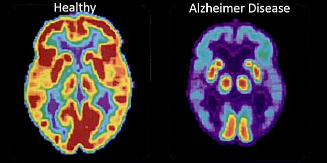
digging around looking at this data

Found this study


Found this study
PET scans accurately identify amyloid deposition after traumatic brain injury

Image: PD
1. Amyloid plaques, similar to those found in Alzheimer’s disease were found in traumatic brain injury (TBI) victims.
2. PET scans accurately identified amyloid deposition and may help further investigate the sequelae TBI.
Evidence Rating Level: 2 (Good)
Study Rundown: Amyloid plaque deposition, similar to that observed in Alzheimer’s disease (AD), patients has been observed in a portion of traumatic brain injury (TBI) victims. It has been found that the Aβ plaques can precipitate within hours of a TBI in post-mortem studies. Studying plaque deposition may allow for a better understanding the long term consequences of TBI as well as AD.
Pittsburgh compound B (PiB) is widely accepted as a ligand marker for cerebral amyloid deposition in AD patients. The authors analyzed the effectiveness of using [11C]PiB to monitor amyloid deposition in TBI patients by comparing [11C]PiB labeled PET scans of TBI patients with brain autopsy specimens labeled with immunohistochemistry and autoradiography. [11C]PiB was found to be a moderately sensitive but specific marker for amyloid deposition following TBI. The study’s greatest limitations are its small sample size and restriction to autopsy specimens that were acquired at most 70 days after TBI. This precludes both adequate analysis of significance and exploration of long-term plaque formation. The study is, however, the first to demonstrate the possible effectiveness of PET scanning to verify amyloid deposition in TBI victims and may serve as a foundation for future research.
Relevant Reading: Head injury as a risk factor for Alzheimer’s disease
In-Depth: This study compared amyloid deposition, identified by [11C]PiB labeled PET scans in TBI patients, to amyloid deposition, identified by immunohistochemistry and autoradiography, in autopsy specimens from TBI victims. Autopsy specimens were also tested with [3H]PiB to compare in vitro binding to immunohistochemistry. PET scans were conducted on 15 TBI patients at various times post-injury; up to 349 days in one patient. Controls consisted of 11 healthy individuals who were scannedm as well as 16 TBI and 7 non-TBI autopsy specimens used for autoradiography. [11C]PiB was found to bind to amyloid plaques in cortical gray matter and the striatum (p<0.05). However immunohistochemistry revealed samples in which plaques in white matter and the thalamus were not bound to [3H]PiB, suggesting less than optimal sensitivity of PiB. Of note is that no false positives indicating plaque deposition were seen.
By Ravi Shah and Rif Rahman
More from this author: Bystander CPR positively associated with cardiac arrest survival, Terminal cancer patients hold misconceptions of the role of chemotherapy,Home-based walking program an effective treatment for peripheral artery disease, Androgen deprivation therapy increases risk of acute kidney injury in patients with prostate cancer, “Polypill” improves outcomes in cardiovascular disease
©2012-2013 2minutemedicine.com. All rights reserved. No works may be reproduced without expressed written consent from 2minutemedicine.com. Disclaimer: We present factual information directly from peer reviewed medical journals. No post should be construed as medical advice and is not intended as such by the authors, editors, staff or by 2minutemedicine.com. PLEASE SEE A HEALTHCARE PROVIDER IN YOUR AREA IF YOU SEEK MEDICAL ADVICE OF ANY SORT.
No comments:
Post a Comment