Brain’s immune cells linked to Alzheimer’s, Parkinson’s, schizophrenia
Salk and UC San Diego scientists conducted vast microglia survey, revealing links to neurodegenerative diseases and psychiatric illnesses
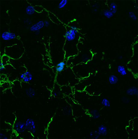
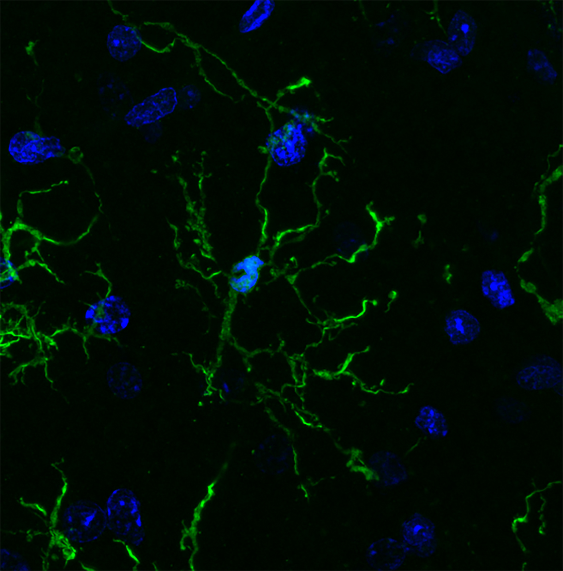
LA JOLLA—Scientists have, for the first time, characterized the molecular markers that make the brain’s front lines of immune defense—cells called microglia—unique. In the process, they discovered further evidence that microglia may play roles in a variety of neurodegenerative and psychiatric illnesses, including Alzheimer’s, Parkinson’s and Huntington’s diseases as well as schizophrenia, autism and depression.
“Microglia are the immune cells of the brain, but how they function in the human brain is not well understood,” says Rusty Gage, professor in Salk’s Laboratory of Genetics, the Vi and John Adler Chair for Research on Age-Related Neurodegenerative Disease, and a senior author of the new work. “Our work not only provides links to diseases but offers a jumping off point to better understand the basic biology of these cells.”
Salk and UC San Diego scientists conducted a vast survey of microglia (pictured here), revealing links to neurodegenerative diseases and psychiatric illnesses.
Genes that have previously been linked to neurological diseases are turned on at higher levels in microglia compared to other brain cells, the team reported in Science on May 25, 2017. While the link between microglia and a number of disorders has been explored in the past, the new study offers a molecular basis for this connection.
“These studies represent the first systematic effort to molecularly decode microglia,” says Christopher Glass, a Professor of Cellular and Molecular Medicine and Professor of Medicine at University of California San Diego, also senior author of the paper. “Our findings provide the foundations for understanding the underlying mechanisms that determine beneficial or pathological functions of these cells.”
Microglia are a type of macrophage, white blood cells found throughout the body that can destroy pathogens or other foreign materials. They’re known to be highly responsive to their surroundings and respond to changes in the brain by releasing pro-inflammatory or anti-inflammatory signals. They also prune back the connections between neurons when cells are damaged or diseased. But microglia are notoriously hard to study. They can’t be easily grown in a culture dish and quickly die outside of a living brain.
Nicole Coufal, a pediatric critical care doctor at UC San Diego, who also works in the Gage lab at Salk, wanted to make microglia from stem cells. But she realized there wasn’t any way to identify whether the resulting cells were truly microglia.
“There was not a unique marker that differentiated microglia from circulating macrophages in the rest of the body,” she says.
David Gosselin and Dylan Skola in the Glass lab, together with Coufal and their collaborators, set out to characterize the molecular characteristics of microglia. They worked with neurosurgeons at UC San Diego to collect brain tissue from 19 patients, all of who were having brain surgery for epilepsy, a brain tumor or a stroke. They isolated microglia from areas of tissue that were unaffected by disease, as well as from mouse brains, and then set out to study the cells. The work was made possible by a multidisciplinary collaboration between bench scientists, bioinformaticians and clinicians.
The team used a variety of molecular and biochemical tests—performed within hours of the cells being collected—to characterize which genes are turned on and off in microglia, how the DNA is marked up by regulatory molecules, and how these patterns change when the cells are cultured.
Microglia, they found, have hundreds of genes that are more highly expressed than other types of macrophages, as well as distinct patterns of gene expression compared to other types of brain cells. After the cells were cultured, however, the gene patterns of the microglia began to change. Within just six hours, more than 2,000 genes had their expression turned down by at least fourfold. The results underscore how dependent microglia are on their surroundings in the brain, and why researchers have struggled to culture them.
From left: Rusty Gage (Salk Institute) and Christopher Glass (UC San Diego).
Next, the researchers analyzed whether any of the genes that were upregulated in microglia compared to other cells had been previously implicated in disease. Genes linked to a variety of neurodegenerative and psychiatric diseases, they found, were highly expressed in microglia.
“A really high proportion of genes linked to multiple sclerosis, Parkinson’s and schizophrenia are much more highly expressed in microglia than the rest of the brain,” says Coufal. “That suggests there’s some kind of link between microglia and the diseases.”
For Alzheimer’s, more than half of the genes known to affect a person’s risk of developing the disease were expressed more highly in microglia than other brain cells.
In mice, however, many of the disease genes weren’t as highly expressed in microglia. “That tells us that maybe mice aren’t the best model organisms for some of these diseases,” Coufal says.
More work is needed to understand exactly how microglia may be altered in people with diseases, but the new molecular profile of microglia offers a way for researchers to begin trying to better culture the cells, or coax stem cells to develop into microglia for future studies.
Other researchers on the study were Baptiste Jaeger, Carolyn O’Connor, Conor Fitzpatrick, Monique Pena, and Amy Adair of the Salk Institute; Inge Holtman, Johannes Schlachetzki, Eniko Sajti, Martina Pasillas, David Gona, and Michael Levy of the University of California San Diego; and Richard Ransohoff of Biogen.
The message from recent surveys is that it's not just people with a diagnosis of schizophrenia who hear voices in their heads, many people considered mentally well do to. This revelation may have a welcome de-stigmatising effect in terms of how people think about some of the symptoms associated with a diagnosis of schizophrenia, but a new study published in Psychosis asks us to hang on a minute – to say that one "hears voices" can mean different things to different people. You might assume that "hears voices" means that a person has an hallucinated auditory experience just like someone is talking to them. But what about hearing an inner voice that is experienced like an out-of-control thought rather than an external voice? Or a heard voice that's not like either a thought or an external voice?
Our knowledge of the experience of voice hearing among patients has been limited by the fact that a lot of psychiatric research in this area (though not all) has been categorical in nature. For instance, a typical psychiatric scale used in research or the clinic includes a vague item like "[Patient] Reports voices than no one else hears" and a tick here can conceal a huge range of different experiences.
For the new research, Nev Jones and Tanya Luhrmann conducted in-depth interviews with 80 people diagnosed with schizophrenia in the US, India and Ghana about their first-hand experiences of hearing voices. There was great variety between participants in their descriptions of voice hearing and also within each individual participant's own descriptions. Perhaps most importantly, while 79 per cent of the participants reported at least some limited experience of the hallucinated sound of external voices – as if someone was audibly speaking to them – a much smaller proportion (17.5 per cent) said this was theirdominant experience of "voice hearing".
The most common dominant experience for the participants was to say they had a mixture of auditory and thought-like voices – 29 per cent reported having this. Another 15 per cent said their dominant experience was to hear thought-like voices that seemed "foreign and alien" but clearly "non-auditory". Other categories were "in-between", being neither auditory or thought-like; "limited auditory" (an auditory experience but just simple words or sounds); "transformed", which is when real voices or sounds were misheard as saying something different; and "multi-sensory", involving visual experiences as much as or more than auditory – for example, one participant described how "the voice will show me all my enemies".
Intriguingly, some participants described how a voice could begin as thought-like, but if they ignored it, then it became more auditory: "If I try ignoring them inside my brain, like they come out. They start telling me things."
The researchers said the interviews showed there is huge variety in the ways that people diagnosed with schizophrenia hear voices, and that it is very difficult for both patients and clinicians to find ways to accurately describe these experiences. Another thing – many of the participants said that what they found most difficult about "hearing voices" was the disruption to their thoughts, rather than the sensory aspect of the experience. Indeed, by emphasising the normality of the auditory aspect of hearing voices, the researchers said there was a risk of mental health professionals presenting a "misleading view of what at least some patients are in fact struggling with".
_________________________________ Jones, N., & Luhrmann, T. (2016). Beyond the sensory: Findings from an in-depth analysis of the phenomenology of “auditory hallucinations” in schizophrenia Psychosis, 8 (3), 191-202 DOI:10.1080/17522439.2015.1100670
Montreal, April 3, 2014 – Bruno Giros, PhD, a researcher at the Douglas Mental Health University Institute and a professor in the Department of Psychiatry at McGill University, has demonstrated, for the first time, the role that dopamine plays in a region of the brain called the hippocampus. Published in Biological Psychiatry, this discovery opens the door to a better understanding of psychiatric diseases, such as schizophrenia.
Dopamine is a neurotransmitter that plays a central role in brain function, and many mental illnesses involve an imbalance in this chemical. What Bruno Giros has shown in particular is that dopamine is present in the hippocampus—the brain area associated with memory and learning—and that it plays a key role in this region.
"Our work helps us better understand some of the symptoms of schizophrenia for which the cause in the brain was unknown, particularly in the area of memory and learning. In a few years, this research could help researchers come up with new therapeutic approaches to improve these symptoms," explained Bruno Giros.
Dr. Giros is the Graham Boeckh Chair in Schizophrenia and the Canada Research Chair in Neurobiology of Mental Disorders. He is one of the world's leading scientists in the study and treatment of schizophrenia. In 1999, he created the Neurobiology and Psychiatry Laboratory at the Institut national de la santé et de la recherche médicale (INSERM) in France. He came to the Douglas Institute in 2008.
###
For information and interviews:
Florence Meney Media Relations Communications and Public Affairs Directorate Douglas Mental Health University Institute Dobell Pav.- 6875 LaSalle Blvd., B-2122 - Montreal, QC H4H 1R3 T. 514-761-6131, ext. 2769 Florence.meney@douglas.mcgill.ca
The Douglas is a world-class institute affiliated with McGill University and the World Health Organization. It treats people suffering from mental illness and offers them both hope and healing. Its teams of specialists and researchers are constantly increasing scientific knowledge, integrating this knowledge into patient care, and sharing it with the community in order to educate the public and eliminate prejudices surrounding mental health.
AAAS and EurekAlert! are not responsible for the accuracy of news releases posted to EurekAlert! by contributing institutions or for the use of any information through the EurekAlert! system.
People with schizophrenia often misinterpret what they see and experience in the world. New research provides insight into the brain mechanisms that might be responsible for this misinterpretation. The study from the Montreal Neurological Institute and Hospital – The Neuro - at McGill University and McGill University Health Centre, reveals that certain errors in visual perception in people with schizophrenia are consistent with interference or ‘noise’ in a brain signal known as a corollary discharge. Corollary discharges are found throughout the animal kingdom, from bugs to fish to humans, and they are thought to be crucial for monitoring one’s own actions. The study, published in the April 2 issue of the Journal of Neuroscience, identifies a corollary discharge dysfunction in schizophrenia, which could aid with diagnosis and treatment of this difficult disorder. It was carried out in collaboration with researchers Veronica Whitford, Gillian O’Driscoll, and Debra Titone in the Department of Psychology, McGill University.
“A corollary discharge is a copy of a nervous system message that is sent to other parts of the brain, in order to make us aware that we are doing something,” said Dr. Christopher Pack, neuroscientist at The Neuro and lead investigator on the study. “For example, if we want to move our arm, the motor area of the brain sends a signal to the muscles to produce a movement. A copy of this command, which is the corollary discharge, is sent to other regions of the brain, to inform them of the impending movement. If you were moving your arm, and you didn’t have the corollary discharge signal, you might assume that someone else was moving your arm. Similarly, if you generated a thought, and you had an impaired corollary discharge, then you might assume that someone else placed the thought in your mind. Corollary discharges ensure that different areas of the brain are communicating with each other, so that we are aware that we are moving our own arm, talking, or thinking our own thoughts.”
Schizophrenia is a disorder that interferes with the ability to think clearly and to manage emotions. People with schizophrenia often attribute their own thoughts and actions to external sources, as in the case of auditory hallucinations. Other common symptoms include delusions and disorganized thinking and speech.
Recent research has suggested that an impaired corollary discharge can account for some of these symptoms. However, the nature of the impairment was unknown. In their study, Dr. Pack and his colleagues (including Dr. Alby Richard, neurology resident at The Neuro) used a test called a perisaccadic localization task, to investigate corollary discharge activity. In this test, subjects are asked to make quick eye movements to follow a dot on a computer screen. At the same time they are also asked to localize visual stimuli that appear briefly on the screen from time to time. In order to perform this task accurately, subjects need to know where on the screen they are looking – in other words they use corollary discharges signals that arise from the brain structures that control the eye muscles.
The results showed that people with schizophrenia were less accurate in figuring out where they were looking. Consequently they made more mistakes in estimating the position of the stimuli that were flashed on the screen. “What is interesting and potentially clinically important is that the pattern of mistakes made by the patients correlated with the extent of their symptoms,” said Dr. Pack. ““This is particularly interesting because the circuits that control eye movements include the best-understood structures in the brain. So we are optimistic that we can work backward from the behavioral data to the biological basis of the corollary discharge effects. We have already started to do this with computational modeling. Mathematically we can convert the corollary discharge of a healthy control into the corollary discharge of a patient with schizophrenia by adding noise and randomness. It is not that people with schizophrenia have no corollary discharge, or a corollary discharge with delayed or weaker amplitude. Rather the patients appear primarily to have a noisy corollary discharge signal. This visual test is very easy thing to do and quite sensitive to individual differences. “
The study shows that patients with schizophrenia make larger errors in localizing visual stimuli compared to controls. These results could be explained by a corollary discharge signal, which also predicts patient symptom severity, suggesting a possible basis for some of the most common symptoms of schizophrenia. This work was supported by The Natural Sciences and Engineering Research Council of Canada, The Brain & Behavior Research Foundation (NARSAD) and the EJLB Foundation.
The Neuro
The Montreal Neurological Institute and Hospital — The Neuro, is a unique academic medical centre dedicated to neuroscience. Founded in 1934 by the renowned Dr. Wilder Penfield, The Neuro is recognized internationally for integrating research, compassionate patient care and advanced training, all key to advances in science and medicine. The Neuro is a research and teaching institute of McGill University and forms the basis for the Neuroscience Mission of the McGill University Health Centre. Neuro researchers are world leaders in cellular and molecular neuroscience, brain imaging, cognitive neuroscience and the study and treatment of epilepsy, multiple sclerosis and neuromuscular disorders. For more information, visit theneuro.com.
Being left-handed has been linked to many mental disorders, but Yale researcher Jadon Webb and his colleagues have found that among those with mental illnesses, people with psychotic disorders like schizophrenia are much more likely to be left-handed than those with mood disorders like depression or bipolar syndrome.
The new study is published in the October-December 2013 issue of the journal SAGE Open.
About 10% of the U.S. population is left-handed. When comparing all patients with mental disorders, the research team found that 11% of those diagnosed with mood disorders such as depression and bipolar disorder are left-handed, which is similar to the rate in the general population. But according to Webb, a child and adolescent psychiatry fellow at the Yale Child Study Center with a particular interest in biomarkers of psychosis, "a striking of 40% of those with schizophrenia or schizoaffective disorder are left-handed."
"In general, people with psychosis are those who have lost touch with reality in some way, through hallucinations, delusions, or false beliefs, and it is notable that this symptom constellation seems to correlate with being left-handed," said Webb. "Finding biomarkers such as this can hopefully enable us to identify and differentiate mental disorders earlier, and perhaps one day tailor treatment in more effective ways."
Webb and his colleagues studied 107 individuals from a public outpatient psychiatric clinic seeking treatment in an urban, low-income community. The research team determined the frequency of left-handedness within the group of patients identified with different types of mental disorders.
The study showed that white patients with psychotic illness were more likely to be left-handed than black patients. "Even after controlling for this, however, a large difference between psychotic and mood disorder patients remained," said Webb.
What sets this study apart from other handedness research is the simplicity of the questionnaire and analysis, said Webb. Patients who were attending their usual check-ups at the mental health facility were simply asked "What hand do you write with?"
"This told us much of what we needed to know in a very simple, practical way," said Webb. "Doing a simple analysis meant that there were no obstacles to participating and we had a very high participation rate of 97%. Patients dealing with serious symptoms of psychosis might have had a harder time participating in a more complicated set of questions or tests. By keeping the survey simple, we were able to get an accurate snapshot of a hard-to-study subgroup of mentally ill people — those who are often poverty-stricken with very poor family and community support."
###
Other authors on the study include Mary I. Schroeder, Christopher Chee, Deanna Dial, Rebecca Hana, Hussam Jefee, Jacob Mays, and Patrick Molitor.
AAAS and EurekAlert! are not responsible for the accuracy of news releases posted to EurekAlert! by contributing institutions or for the use of any information through the EurekAlert! system.