Showing posts with label alzheimer's. Show all posts

Thursday, June 23, 2022

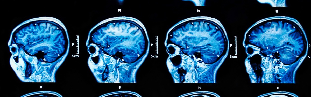
Algorithm could diagnose Alzheimer’s disease from a single brain scan

 reposted from


Algorithm could diagnose Alzheimer’s disease from a single brain scan

Published: 20 June 2022

A single MRI scan of the brain could be enough to diagnose Alzheimer’s disease, according to new research supported by NIHR.

Researchers developed an algorithm to analyse structural features shown on brain MRI scans, including in regions not previously associated with Alzheimer’s. This machine learning technology was able to accurately predict the existence of Alzheimer’s disease and identify the disease at an early stage, when it can be very difficult to diagnose.

Alzheimer’s disease is the most common form of dementia, affecting over half a million people in the UK. Although most people with Alzheimer’s disease develop it after the age of 65, people under this age can develop it too. The most frequent symptoms of dementia are memory loss and difficulties with thinking, problem solving and language.

Currently lots of tests are used to diagnose Alzheimer’s disease, including memory and cognitive tests and brain scans. The scans are used to check for protein deposits in the brain and shrinkage of the hippocampus, the area of the brain linked to memory. All of these tests can take several weeks, both to arrange and to process.

Getting a diagnosis quickly at an early stage helps patients access help and support, get treatment to manage their symptoms, and plan for the future. Being able to accurately identify patients at an early stage of the disease will also help researchers to understand the brain changes that trigger Alzheimer’s disease, and support development and trials of new treatments.

The researchers, supported by Imperial Biomedical Research Centre, studied just one of the tests currently used to diagnose Alzheimer’s disease - an MRI scan. They adapted an algorithm developed for use in classifying cancer tumours and applied it to MRI scans of the brain.

The researchers divided the brain into 115 regions and allocated 660 different features, such as size, shape and texture. They then trained the algorithm to identify where changes to these features could accurately predict the existence of Alzheimer’s disease.

Using data from the Alzheimer’s Disease Neuroimaging Initiative, the team tested their approach on brain scans from over 400 patients with early and later stage Alzheimer’s, healthy controls and patients with other neurological conditions, including frontotemporal dementia and Parkinson’s disease. They also tested it with data from more than 80 patients undergoing diagnostic tests for Alzheimer’s at Imperial College Healthcare NHS Trust.

The research, published in the Nature Portfolio Journal Communications Medicine, found that in 98% of cases, the MRI-based machine learning system alone could accurately predict whether the patient had Alzheimer’s disease or not. It was also able to distinguish between early and late-stage Alzheimer’s with fairly high accuracy, in 79% of patients.

The new system spotted changes in areas of the brain not previously associated with Alzheimer’s disease, including the cerebellum (the part of the brain that coordinates and regulates physical activity) and the ventral diencephalon (linked to the senses, sight and hearing). This opens up potential new avenues for research into these areas and their links to Alzheimer’s disease.

Professor Eric Aboagye, from Imperial’s Department of Surgery and Cancer, who led the research, said: “Currently no other simple and widely available methods can predict Alzheimer’s disease with this level of accuracy, so our research is an important step forward. Many patients who present with Alzheimer’s at memory clinics do also have other neurological conditions, but even within this group our system could pick out those patients who had Alzheimer’s from those who did not.

“Waiting for a diagnosis can be a horrible experience for patients and their families. If we could cut down the amount of time they have to wait, make diagnosis a simpler process, and reduce some of the uncertainty, that would help a great deal. Our new approach could also identify early-stage patients for clinical trials of new drug treatments or lifestyle changes, which is currently very hard to do.”

Dr Paresh Malhotra, who is a consultant neurologist at Imperial College Healthcare NHS Trust and a researcher in Imperial’s Department of Brain Sciences, said: “Although neuroradiologists already interpret MRI scans to help diagnose Alzheimer’s, there are likely to be features of the scans that aren’t visible, even to specialists. Using an algorithm able to select texture and subtle structural features in the brain that are affected by Alzheimer’s could really enhance the information we can gain from standard imaging techniques.”

Read more about this research on the NIHR imperial BRC website

Monday, November 16, 2020

New Tracer Gives Clear Picture of Alzheimer’s and Other Dementias

 reposted from The Scientist


New Tracer Gives Clear Picture of Alzheimer’s and Other Dementias

New Tracer Gives Clear Picture of Alzheimer’s and Other Dementias

An imaging agent reveals aggregated tau protein in the brain during PET scans and could improve the diagnosis of neurodegenerative diseases, particularly tauopathies.

Ian Le Guillou
Ian Le Guillou
Oct 29, 2020
118

ABOVE: PET scans of individuals using the new tau tracer
TAGAI, ONO, AND KUBOTA ET AL.

Anew tracer for brain imaging could offer a clear window into the development of Alzheimer’s disease and other neurodegenerative conditions. The tracing agent, which highlights the accumulation of toxic tau protein deposits in the brain, could distinguish between a range of conditions called tauopathies that can be difficult to tell apart at the early stages, such as frontotemporal dementia and progressive supranuclear palsy.

Tau is a protein involved in maintaining the structure of neurons. In tauopathies, including Alzheimer’s disease, it aggregates to form knots inside the cells, eventually killing them. 

PET scans are a common diagnostic technique used in hospitals, relying on a radioactive tracing agent to reveal the location of a molecule of interest. Earlier this year, the US Food and Drug Administration approved the first tau PET tracer for Alzheimer’s disease, Tauvid. It has proved challenging to find reliable PET tracers that can bind to the various forms of tau seen in different tauopathies. 

In pursuit of a tau tracer that would offer a sharper signal than previous iterations in development, researchers adapted an existing tracer to make it last longer in the body. The new tracer, known as 18F-PM-PBB3, carries a radioactive fluorine isotope to the tau tangles and releases a positron particle detected by a PET scanner when it binds its target.

While the developers succeeded in enhancing tau visualization, their tracer also had the unexpected effect of binding to a greater variety of tau deposits seen across different tauopathies. This means that the same tracer could be used as a diagnostic test for multiple neurodegenerative conditions.

The researchers tested the tracer in 39 patients with a range of tau-related conditions, including Alzheimer’s disease. Based on the location of the tracer’s signal on the brain scans, the team was able to predict accurately the type of disease, which was confirmed in some cases by later autopsies. Their results appear today (October 29) in Neuron.

In the development of Alzheimer’s disease, the tau deposits are thought to follow on from the accumulation of amyloid-β plaques. Although most efforts to create treatments for Alzheimer’s have focused on amyloid, the presence of tau is thought to be more closely linked to the development of symptoms. The researchers were able to accurately predict the severity of the symptoms of Alzheimer’s disease in 17 people based on the abundance of tau seen in the scans.

“Even during the clinical trials for anti-amyloid drugs, we also need to pursue the tau accumulation in those patients to see whether or not the tau accumulation, which is closely associated with the neuronal death, could be suppressed as a result of the amyloid-β suppression,” says coauthor Makoto Higuchi of the National Institute of Radiological Sciences in Japan.

The tracer may have the biggest effect on diseases other than Alzheimer’s. Experimental tracers that had previously been developed were unable to detect all forms of tau seen in tauopathies. “One of the problems with the old tracers was that the tau pathology in Alzheimer’s disease could be detected, but there were problems with detecting it in the other tauopathies. So this opens new perspectives. Now we really can image the pathology in the patients, and we can make the link with the symptoms,” says Ilse Dewachter, a neuroscientist at Hasselt University in Belgium who was not involved in the study. 

In conditions such as frontotemporal dementia, which can be caused by tau, the early symptoms can be significant behavioral changes and a loss of inhibition, leading to social problems. “Even without the radical cure for the tauopathy, we can socially assist those people by predicting the emerging, symptomatic problems in each of the subjects. That’s the major advantage of imaging tau deposits in the brain,” says Higuchi.

PET scans would be too expensive for use in mass screening for dementia, but Higuchi notes that they could still be helpful in developing tests that instead look for biomarkers in the blood or cerebrospinal fluid. As part of such an effort, scans would first categorize individuals as being tau-positive or tau-negative, and then researchers could identify biomarkers that distinguish between the two groups.

See “The Hunt for a Blood Test for Alzheimer’s Disease”

The imaging might also help identify the right people to take part in clinical trials, as proteins other than tau can cause frontotemporal dementia.

“If you want to be able to develop trials for patients who present with the same syndromes, we need to know what subtype of pathology they have. So far, we have no biomarker. It’s really, really hard right now to guess if it’s tau or another [protein aggregating],” says Renaud La Joie, a neuroscientist at the University of California, San Francisco, who was not involved in the study. “For non-Alzheimer’s trials, we really need help screening the right patients.” 

Higuchi and his colleagues have patented the tracer and licensed it to APRINOIA Therapeutics, which is testing its use as a diagnostic tool for Alzheimer’s disease in clinical trials in the US, China, and Japan. Higuchi says he hopes that they will soon be able to do clinical trials for diagnosing other forms of dementia.

K. Tagai et al., “High-contrast in vivo imaging of tau pathologies in Alzheimer’s and non-Alzheimer’s disease tauopathies,” Neuron,  doi:10.1016/j.neuron.2020.09.042, 2020.

Tuesday, March 26, 2019

Biomarkers of Alzheimer’s Disease

reposted from
https://sapienlabs.co/biomarkers-of-alzheimers-disease/


Biomarkers of Alzheimer’s Disease

Alzheimer’s disease has very specific etiology that can typically only be confirmed postmortem. Are there ways to identify it in the dynamical features of brain activity?
Alzheimer’s disease (AD), a neurodegenerative disorder characterized by a decline in cognitive functioning, in particular memory loss, is the most common cause of dementia with an estimated 30 million people affected worldwide [1,2].  At a neurobiological level it is characterized by aggregations of beta-amyloid (Aβ) protein into plaques, the accumulation of tau protein neurofibrillary tangles and progressive neurodegeneration. One recent question of interest is how these structural changes translate into changes in brain activity. Can it be reliably measured in the EEG to provide biomarkers of disease onset and progression, allowing clinicians to make an early diagnosis and intervention?

Biomarkers for early intervention.

For most of its history, AD has been diagnosed solely through clinical observation and cognitive testing, with a confirmatory diagnosis only performed on postmortem examination. However, the neurobiological changes associated with AD, and a potential precursor, Mild Cognitive Impairment (MCI), often appear many years (or even decades) before any visible clinical signs in the patient.
The advent of neuroimaging and the development of new biomarkers offer clinicians the opportunity to do this [3,4]. However, the challenges of developing either structurally or functionally relevant AD biomarkers which provide accurate and reliable indicators of disease onset, progression and outcome, or which assist in drug development, are considerable.
Examples of currently accepted biomarkers involve measuring levels of brain chemicals related to amyloid or tau (e.g. in the cerebrospinal fluid, CSF), or through estimates of metabolic activity (e.g. with Positron Emission Tomography, PET). For example, CSF levels of amyloid-beta (Aβ42) and phosphorylated tau (p-Tau) are thought to reflect AD pathology. In addition, the formation of plaques and tangles disrupt the balance of excitatory and inhibitory activity in the brain, and also result in synaptic dysfunction, at least in mouse models [5], both of which affect brain dynamics.  This provides an opportunity for studying the progression of AD with techniques such as resting-state EEG.

LORETA and Alzheimer’s Disease.

Multiple studies have attempted to examine changes in resting-state EEG dynamics, and to relate these to other markers of AD [6]. For example, in one recent small-scale study, resting-state EEG was used to explore whether there was a relationship between cortical hypometabolism – something commonly observed in AD – and cortical EEG rhythms [7]. To do this they measured cortical hypometabolism using fluorodeoxyglucose-PET and recorded resting EEG in 19 AD patients and compared this against 40 healthy controls and analyzed the results using LORETA. The EEG results showed higher levels of source localized delta band activity that correlated (r=0.579, p=0.009, N=19) with measures of cortical hypometabolism (other bands were not statistically different). This suggests that, in AD patients, delta activity at rest may be related to the PET biomarker of cortical hypometabolism. However, since the healthy patients did not agree to a PET scan, it limits the validity of this conclusion.  Also, such conclusion is confounded by similar results relating to a host of other mental health disorders and may simply be representative of a disorder in general, but not AD specifically.
Grand average across subjects of the normalized LORETA solutions. From [6]

CSF markers and Alzheimer’s disease

Another larger-scale study explored the relationship between EEG measures and CSF biomarkers [8]. In this study they compared patients with subjective cognitive decline (n=210) (i.e. they reported subjective complaints but had no significant cognitive deficit or clinical symptoms) against those with MCI (n=230) or AD (n=197). They analyzed resting-state EEG data using two different metrics – global field power (GFP) and global field synchronization (GFS). GFP is a reference-free method that reduces multichannel recordings to a single measure corresponding to the generalized EEG amplitude, resulting in a global measure of scalp potential field strength whilst GFS is a measure of global functional connectivity which resembles the global amount of instantaneous phase locked synchronization of oscillating neuronal networks across the scalp. Linear regression models showed that decreased levels of Aβ42 in the CSF significantly correlated with increased theta (β coefficient=0.514, p<0.001) and delta (β coefficient=0.304, p=0.001) GFP. In addition, decreased levels of Aβ42 in the CSF were significantly associated with decreased GFS alpha (β coefficient=0.024, p<0.001). and beta (β coefficient=0.013, p<0.001). These latter correlations were present in individuals with subjective cognitive decline, suggesting that GFS may be a potential pre-clinical marker of early AD.

Integrative Biomarkers

These two studies provide a snapshot into the direction of research and progress that is being made in the development of potential resting-state EEG biomarkers which track the progression of AD (other research focuses on task related ERP biomarkers which isn’t discussed here). However, it is unlikely that a single biomarker will be sufficient in adequately predicting the onset, progression and outcome of AD. One longitudinal study which has tried to address this monitored 86 patients initially diagnosed with MCI over a period of 2 years [9]. During this time 25 of the patients developed AD allowing them to search for a marker indicating the likelihood of a patient converting from MCI to AD. They measured multiple different biomarkers and found that several EEG biomarkers based around the alpha and beta range were associated with the conversion from MCI to AD. Rather than focusing on just one of these, they found that by integrating 6 of them together they were able to develop a diagnostic tool that predicted AD progression with a sensitivity of 88% and specificity of 82%. This was compared to a sensitivity of 64% and specificity of 62% when only a single biomarker was used.
The 6 Biomarkers of Interest. From [9]

The Verdict

EEG offers an opportunity to support the early identification of Alzheimer’s disease and so far there are promising directions. However as with many EEG biomarkers, this search is also hindered by inconsistencies in the methodological approach across studies [6].  More significantly, there is a substantial challenge of identifying markers that are specific to AD and not general to all cognitive and mental health function, one that may be  overcome by studying the EEG in multiple forms of Dementia together in combination with multiple other types of markers.

References:
[1] McKhann, G., Knopman, D., Chertkow, H., Hyman, B., Jack, C., & Kawas, C. et al. (2011). The diagnosis of dementia due to Alzheimer’s disease: Recommendations from the National Institute on Aging-Alzheimer’s Association workgroups on diagnostic guidelines for Alzheimer’s disease. Alzheimer’s & Dementia, 7(3), 263-269. doi: 10.1016/j.jalz.2011.03.005
[2] Holtzman, D., Morris, J., & Goate, A. (2011). Alzheimer’s Disease: The Challenge of the Second Century. Science Translational Medicine, 3(77), 77sr1-77sr1. doi: 10.1126/scitranslmed.3002369
[3] Maestú, F., Cuesta, P., Hasan, O., Fernandéz, A., Funke, M., & Schulz, P. (2019). The Importance of the Validation of M/EEG With Current Biomarkers in Alzheimer’s Disease. Frontiers In Human Neuroscience, 13. doi: 10.3389/fnhum.2019.00017
[4] Wurtman, R. (2015). Biomarkers in the diagnosis and management of Alzheimer’s disease. Metabolism, 64(3), S47-S50. doi: 10.1016/j.metabol.2014.10.034
[5] Selkoe, D. (2002). Alzheimer’s Disease Is a Synaptic Failure. Science, 298(5594), 789-791. doi: 10.1126/science.1074069
[6] Cassani, R., Estarellas, M., San-Martin, R., Fraga, F., & Falk, T. (2018). Systematic Review on Resting-State EEG for Alzheimer’s Disease Diagnosis and Progression Assessment. Disease Markers, 2018, 1-26. doi: 10.1155/2018/5174815
[7] Babiloni, C., Del Percio, C., Caroli, A., Salvatore, E., Nicolai, E., & Marzano, N. et al. (2016). Cortical sources of resting state EEG rhythms are related to brain hypometabolism in subjects with Alzheimer’s disease: an EEG-PET study. Neurobiology Of Aging, 48, 122-134. doi: 10.1016/j.neurobiolaging.2016.08.021
[8] Smailovic, U., Koenig, T., Kåreholt, I., Andersson, T., Kramberger, M., Winblad, B., & Jelic, V. (2018). Quantitative EEG power and synchronization correlate with Alzheimer’s disease CSF biomarkers. Neurobiology Of Aging, 63, 88-95. doi: 10.1016/j.neurobiolaging.2017.11.005
[9] Poil, S., de Haan, W., van der Flier, W., Mansvelder, H., Scheltens, P., & Linkenkaer-Hansen, K. (2013). Integrative EEG biomarkers predict progression to Alzheimer’s disease at the MCI stage. Frontiers In Aging Neuroscience, 5. doi: 10.3389/fnagi.2013.00058

Thursday, March 7, 2019

Germs in Your Gut Are Talking to Your Brain. Scientists Want to Know What They’re Saying

reposted from


MATTER

Germs in Your Gut Are Talking to Your Brain. Scientists Want to Know What They’re Saying.

The body’s microbial community may influence the brain and behavior, perhaps even playing a role in dementia, autism and other disorders.
CreditSean McSorley
Image
CreditSean McSorley
In 2014 John Cryan, a professor at University College Cork in Ireland, attended a meeting in California about Alzheimer’s disease. He wasn’t an expert on dementia. Instead, he studied the microbiome, the trillions of microbes inside the healthy human body.
Dr. Cryan and other scientists were beginning to find hints that these microbes could influence the brain and behavior. Perhaps, he told the scientific gathering, the microbiome has a role in the development of Alzheimer’s disease.
The idea was not well received. “I’ve never given a talk to so many people who didn’t believe what I was saying,” Dr. Cryan recalled.
A lot has changed since then: Research continues to turn up remarkable links between the microbiome and the brain. Scientists are finding evidence that microbiome may play a role not just in Alzheimer’s disease, but Parkinson’s disease, depression, schizophrenia, autism and other conditions.
For some neuroscientists, new studies have changed the way they think about the brain.
One of the skeptics at that Alzheimer’s meeting was Sangram Sisodia, a neurobiologist at the University of Chicago. He wasn’t swayed by Dr. Cryan’s talk, but later he decided to put the idea to a simple test.
“It was just on a lark,” said Dr. Sisodia. “We had no idea how it would turn out.”
He and his colleagues gave antibiotics to mice prone to develop a version of Alzheimer’s disease, in order to kill off much of the gut bacteria in the mice. Later, when the scientists inspected the animals’ brains, they found far fewer of the protein clumps linked to dementia.
Just a little disruption of the microbiome was enough to produce this effect. Young mice given antibiotics for a week had fewer clumps in their brains when they grew old, too.
“I never imagined it would be such a striking result,” Dr. Sisodia said. “For someone with a background in molecular biology and neuroscience, this is like going into outer space.”
Following a string of similar experiments, he now suspects that just a few species in the gut — perhaps even one — influence the course of Alzheimer’s disease, perhaps by releasing chemical that alters how immune cells work in the brain.
He hasn’t found those microbes, let alone that chemical. But “there’s something’s in there,” he said. “And we have to figure out what it is.”
Scientists have long known that microbes live inside us. In 1683, the Dutch scientist Antonie van Leeuwenhoek put plaque from his teeth under a microscope and discovered tiny creatures swimming about.
But the microbiome has stubbornly resisted scientific discovery. For generations, microbiologists only studied the species that they could grow in the lab. Most of our interior occupants can’t survive in petri dishes.
In the early 2000s, however, the science of the microbiome took a sudden leap forward when researchers figured out how to sequence DNA from these microbes. Researchers initially used this new technology to examine how the microbiome influences parts of our bodies rife with bacteria, such as the gut and the skin.
Few of them gave much thought to the brain — there didn’t seem to be much point. The brain is shielded from microbial invasion by the so-called blood-brain barrier. Normally, only small molecules pass through.
“As recently as 2011, it was considered crazy to look for associations between the microbiome and behavior,” said Rob Knight, a microbiologist at the University of California, San Diego.
He and his colleagues discovered some of the earliest hints of these links. Investigators took stool from mice with a genetic mutation that caused them to eat a lot and put on weight. They transferred the stool to mice that had been raised germ-free — that is, entirely without gut microbiomes — since birth.
After receiving this so-called fecal transplant, the germ-free mice got hungry, too, and put on weight.
Altering appetite isn’t the only thing that the microbiome can do to the brain, it turns out. Dr. Cryan and his colleagues, for example, have found that mice without microbiomes become loners, preferring to stay away from fellow rodents.
The scientists eventually discovered changes in the brains of these antisocial mice. One region, called the amygdala, is important for processing social emotions. In germ-free mice, the neurons in the amygdala make unusual sets of proteins, changing the connections they make with other cells.
Studies of humans revealed some surprising patterns, too. Children with autism have unusual patterns of microbial species in their stool. Differences in the gut bacteria of people with a host of other brain-based conditions also have been reported.
But none of these associations proves cause and effect. Finding an unusual microbiome in people with Alzheimer’s doesn’t mean that the bacteria drive the disease. It could be the reverse: People with Alzheimer’s disease often change their eating habits, for example, and that switch might favor different species of gut microbes.
Fecal transplants can help pin down these links. In his research on Alzheimer’s, Dr. Sisodia and his colleagues transferred stool from ordinary mice into the mice they had treated with antibiotics. Once their microbiomes were restored, the antibiotic-treated mice started developing protein clumps again.
“We’re extremely confident that it’s the bacteria that’s driving this,” he said. Other researchers have taken these experiments a step further by using human fecal transplants.
If you hold a mouse by its tail, it normally wriggles in an effort to escape. If you give it a fecal transplant from humans with major depression, you get a completely different result: The mice give up sooner, simply hanging motionless.
As intriguing as this sort of research can be, it has a major limitation. Because researchers are transferring hundreds of bacterial species at once, the experiments can’t reveal which in particular are responsible for changing the brain.
Now researchers are pinpointing individual strains that seem to have an effect.
To study autism, Dr. Mauro Costa-Mattioli and his colleagues at the Baylor College of Medicine in Houston investigated different kinds of mice, each of which display some symptoms of autism. A mutation in a gene called SHANK3 can cause mice to groom themselves repetitively and avoid contact with other mice, for example.
In another mouse strain, Dr. Costa-Mattioli found that feeding mothers a high-fat diet makes it more likely their pups will behave this way.
When the researchers investigated the microbiomes of these mice, they found the animals lacked a common species called Lactobacillus reuteri. When they added a strain of that bacteria to the diet, the animals became social again.
Dr. Costa-Mattioli found evidence that L. reuteri releases compounds that send a signal to nerve endings in the intestines. The vagus nerve sends these signals from the gut to the brain, where they alter production of a hormone called oxytocin that promotes social bonds.
Other microbial species also send signals along the vagus nerve, it turns out. Still others communicate with the brain via the bloodstream.
It’s likely that this influence begins before birth, as a pregnant mother’s microbiome releases molecules that make their way into the fetal brain.
Mothers seed their babies with microbes during childbirth and breast feeding. During the first few years of life, both the brain and the microbiome rapidly mature.
To understand the microbiome’s influence on the developing brain, Rebecca Knickmeyer, a neuroscientist at Michigan State University, is studying fMRI scans of infants.
In her first study, published in January, she focused on the amygdala, the emotion-processing region of the brain that Dr. Cryan and others have found to be altered in germ-free mice.
Dr. Knickmeyer and her colleagues measured the strength of the connections between the amygdala and other regions of the brain. Babies with a lower diversity of species in their guts have stronger connections, the researchers found.
Does that mean a low-diversity microbiome makes babies more fearful of others? It’s not possible to say yet — but Dr. Knickmeyer hopes to find out by running more studies on babies.
CreditSean McSorley
Image
CreditSean McSorley
As researchers better understand how the microbiome influences the brain, they hope doctors will be able to use it to treat psychiatric and neurological conditions.
It’s possible they’ve been doing it for a long time — without knowing.
In the early 1900s, neurologists found that putting people with epilepsy on a diet low in carbohydrates and high in protein and fat sometimes reduced their seizures.
Epileptic mice experience the same protection from a so-called ketogenic diet. But no one could say why. Elaine Hsiao, a microbiologist at the University of California, Los Angeles, suspected that the microbiome was the reason.
To test the microbiome’s importance, Dr. Hsiao and her colleagues raised mice free of microbes. When they put the germ-free epileptic mice on a ketogenic diet, they found that the animals got no protection from seizures.
But if they gave the germ-free animals stool from mice on a ketogenic diet, seizures were reduced.
Dr. Hsiao found that two types of gut bacteria in particular thrive in mice on a ketogenic diet. They may provide their hosts with building blocks for neurotransmitters that put a brake on electrical activity in the brain.
It’s conceivable that people with epilepsy wouldn’t need to go on a ketogenic diet to get its benefits — one day, they may just take a pill containing the bacteria that do well on the diet.
Sarkis Mazmanian, a microbiologist at Caltech, and his colleagues have identified a single strain of bacteria that triggers symptoms of Parkinson’s disease in mice. He has started a company that is testing a compound that may block signals that the microbe sends to the vagus nerve.
Dr. Mazmanian and other researchers now must manage a tricky balancing act. On one hand, their experiments have proven remarkably encouraging; on the other, scientists don’t want to encourage the notion that microbiome-based cures for diseases like Parkinson’s are around the corner.
That’s not easy when people can buy probiotics without a prescription, and when some companies are willing to use preliminary research to peddle microbes to treat conditions like depression.
“The science can get mixed up with what the pseudoscientists are doing,” said Dr. Hsiao.
Dr. Costa-Mattioli hopes that L. reuteri some day will help some people with autism, but he warns parents against treating their children with store-bought probiotics. Some strains of L. reuteri alter the behavior of mice, he’s found, and others don’t.
Dr. Costa-Mattioli and his colleagues are still searching for the most effective strain and figuring out the right dose to try on people. “You want to go into a clinical trial with the best weapon, and I’m not sure we have it,” he said.
Katarzyna B. Hooks, a computational biologist at the University of Bordeaux in France, warned that studies like Dr. Costa-Mattioli’s are still unusual. Most of these findings come from research with fecal transplants or germ-free mice — experiments in which it’s especially hard to pinpoint the causes of changes in behavior.
“We have the edges of the puzzle, and we’re now trying to figure out what’s in the picture itself,” she said.
A version of this article appears in print on , on Page D1 of the New York edition with the headline: Beyond a Gut Feeling. Order Reprints | Today’s Paper | Subscribe